Advanced Bioimaging Laboratory
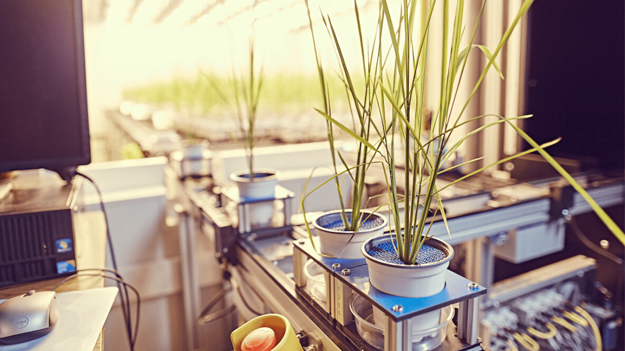
The Donald Danforth Plant Science Center's Advanced Bioimaging Laboratory (ABL) uses state-of-the-art imaging technology to document plants, microbes, and their interactions, from whole plants down to the cellular level. By conducting experiments and using these powerful microscopes to view living organisms, the ABL offers a portal into a world invisible to the naked eye and provides an up-close look at the inner workings of cells and their interactions with their environment. By discovering how plants live and grow through direct observation, the work of the ABL furthers our knowledge of our world and helps us understand how plant science can be used to create a more sustainable future.
2021
Anderson, C.M., Mattoon, E.M., Zhang, N., Becker, E., McHargue, W., Yang, J., Patel, D., Dautermann, O., McAdam, S.A.M., Tarin, T., Pathak, S., Avenson, T.J., Berry, J., Braud, M., Niyogi, K.K., Wilson, M., Nusinow, D.A., Vargas, R., Czymmek, K.J., Eveland, A.L. and Zhang, R. (2021) High light and high temperature reduce photosynthesis via different mechanisms in the c4 model Setaria viridis. bioRxiv, 431694. doi:10.1101/2021.02.20.431694
Cox, K.L., Manchego, J., Meyers, B.C., Czymmek, K.J. and Harkess, A. (2021) Automated imaging of duckweed growth and development. bioRxiv, 453240. doi:10.1101/2021.07.21.453240
Gillman, J.D., Chebrolu, K. and Smith, J.R. (2021) Quantitative trait locus mapping for resistance to heat-induced seed degradation and low seed phytic acid in soybean. Crop Sci 61, 2023-2035. doi:10.1002/csc2.20419
Gurazada, S.G.R., Cox, K.L., Czymmek, K.J. and Meyers, B.C. (2021) Space: the final frontier - achieving single-cell, spatially resolved transcriptomics in plants. Emerg Top Life Sci 5, 179-188. doi:10.1042/etls20200274
Harkess, A., McLoughlin, F., Bilkey, N., Elliott, K., Emenecker, R., Mattoon, E., Miller, K., Czymmek, K., Vierstra, R., Meyers, B.C. and Michael, T.P. (2021) Improved Spirodela polyrhiza genome and proteomic analyses reveal a conserved chromosomal structure with high abundances of chloroplastic proteins favoring energy production. J Exp Bot 72, 2491-2500. doi:10.1101/2020.01.23.909457
Hung, Y.-H. and Slotkin, R.K. (2021) The initiation of RNA interference (RNAi) in plants. Curr Opin Plant Biol 61, 102014. doi:10.1016/j.pbi.2021.102014
Kim, H.S., Kim, J.E., Hwangbo, A., Akerboom, J., Looger, L.L., Duncan, R., Son, H., Czymmek, K.J. and Kang, S. (2021) Evaluation of multi-color genetically encoded Ca(2+) indicators in filamentous fungi. Fungal Genet Biol 149, 103540. doi:10.1016/j.fgb.2021.103540
Romsdahl, T.B., Kambhampati, S., Koley, S., Yadav, U.P., Alonso, A.P., Allen, D.K. and Chapman, K.D. (2021) Analyzing mass spectrometry imaging data of (13)C-labeled phospholipids in Camelina sativa and Thlaspi arvense (Pennycress) embryos. Metabolites 11, 148. doi:10.3390/metabo11030148
Zhang, N., Mattoon, E.M., McHargue, W., Venn, B., Zimmer, D., Pecani, K., Jeong, J., Anderson, C.M., Chen, C., Berry, J.C., Xia, M., Tzeng, S.-C., Becker, E., Pazouki, L., Evans, B., Cross, F., Cheng, J., Czymmek, K.J., Schroda, M., Mühlhaus, T. and Zhang, R. (2021) Systems-wide analysis revealed shared and unique responses to moderate and acute high temperatures in the green alga Chlamydomonas reinhardtii. bioRxiv, 456552. doi:10.1101/2021.08.17.456552
2020
AuBuchon-Elder, T., Coneva, V., Goad, D.M., Jenkins, L.M., Yu, Y., Allen, D.K. and Kellogg, E.A. (2020) Sterile spikelets contribute to yield in sorghum and related grasses. Pl Cell 32, 3500-3518. doi:10.1105/tpc.20.00424
Bélanger, S., Pokhrel, S., Czymmek, K.J. and Meyers, B.C. (2020) Pre-meiotic, 24-nucleotide reproductive phasiRNAs are abundant in anthers of wheat and barley but not rice and maize. Plant Physiol 184, 1407-1423. doi:10.1104/pp.20.00816
Chu, K., Jenkins, L.M., Bailey, S.R., Kambhampati, S., Koley, S., Foley, K., Arp, J.J., Czymmek, K.J., Bates, P.D. and Allen, D.K. (2020) Shifting carbon flux from non-transient starch to lipid allows oil accumulation in transgenic tobacco leaves. bioRxiv, 098632. doi:10.1101/2020.05.15.098632
Czymmek, K., Sawant, A., Goodman, K., Pennington, J., Pedersen, P., Hoon, M. and Otegui, M.S. (2020) Imaging plant cells by high-pressure freezing and serial block-face scanning electron microscopy. Methods Mol Biol 2177, 69-81. doi:10.1007/978-1-0716-0767-1_7
Czymmek, K., Duncan, K., Zhang, N. and Zhang, R. (2020) Strategies for optimizing heavy metal staining in freeze-substituted resin embedded plants samples for electron and x-ray microscopy. Microsc Microanal 26, 142-143. doi:10.1017/S1431927620013550
Duncan, K.E., Czymmek, K.J., Jiang, N., Thies, A.C. and Topp, C.N. (2020) X-ray microscopy enables multiscale high-resolution 3D imaging of plant cells, tissues, and organs. bioRxiv, 423480. doi:10.1101/2020.12.18.423480
Liu, Z., Nguyen, Q.P.H., Nanjundappa, R., Delgehyr, N., Megherbi, A., Doherty, R., Thompson, J., Jackson, C., Albulescu, A., Heng, Y.M., Lucas, J.S., Dell, S.D., Meunier, A., Czymmek, K., Mahjoub, M.R. and Mennella, V. (2020) Super-resolution microscopy and FIB-SEM imaging reveal parental centriole-derived, hybrid cilium in mammalian multiciliated cells. Devel Cell 55, 224-+. doi:10.1016/j.devcel.2020.09.016
Negi, S., Perrine, Z., Friedland, N., Kumar, A., Tokutsu, R., Minagawa, J., Berg, H., Barry, A.N., Govindjee, G. and Sayre, R. (2020) Light-regulation of light harvesting antenna size substantially enhances photosynthetic efficiency and biomass yield in green algae. Plant J 103, 584-603. doi:10.1111/tpj.14751
Prunet, N. and Duncan, K. (2020) Imaging flowers: a guide to current microscopy and tomography techniques to study flower development. J Exp Bot 71, 2898-2909. doi:10.1093/jxb/eraa094
Velivelli, S.L.S., Czymmek, K.J., Li, H., Shaw, J.B., Buchko, G.W. and Shah, D.M. (2020) Antifungal symbiotic peptide NCR044 exhibits unique structure and multifaceted mechanisms of action that confer plant protection. Proc Natl Acad Sci U S A 117, 16043-16054. doi:10.1073/pnas.2003526117
Wu, G.H., Mitchell, P.G., Galaz-Montoya, J.G., Hecksel, C.W., Sontag, E.M., Gangadharan, V., Marshman, J., Mankus, D., Bisher, M.E., Lytton-Jean, A.K.R., Frydman, J., Czymmek, K. and Chiu, W. (2020) Multi-scale 3D cryo-correlative microscopy for vitrified cells. Structure 28, 1231-1237. doi:10.1016/j.str.2020.07.017
2019
Augustin, M.M., Augustin, J.M., Brock, J.R. and Kutchan, T.M. (2019) Enzyme morphinan N-demethylase for more sustainable opiate processing. Nature Sustain 2, 465-474. doi:10.1038/s41893-019-0302-6
Ivanov, S., Austin, J., 2nd, Berg, R.H. and Harrison, M.J. (2019) Extensive membrane systems at the host-arbuscular mycorrhizal fungus interface. Nature Plants 5, 194-203. doi:10.1038/s41477-019-0364-5
Li, H., Velivelli, S.L.S. and Shah, D.M. (2019) Antifungal potency and modes of action of a novel olive tree defensin against closely related ascomycete fungal pathogens. Mol Plant Mic In 32, 1649-1664. doi:10.1094/mpmi-08-19-0224-r
Liberton, M., Bandyopadhyay, A. and Pakrasi, H.B. (2019) Enhanced nitrogen fixation in a glgX-deficient strain of Cyanothece sp. Strain ATCX 51142, a unicellular nitrogen-fixing cyanobacterium. Appl Environ Microbiol 85, e02887-02818. doi:10.1128/aem.02887-18
Singh, R., Low, E.-T.L., Ooi, L.C.-L., Ong-Abdullah, M., Ting, N.-C., Nookiah, R., Ithnin, M., Marjuni, M., Mustaffa, S., Yaakub, Z., Amiruddin, M.D., Manaf, M.A.A., Chan, K.-L., Halim, M.A.A., Sanusi, N.S.N.M., Lakey, N., Sachdeva, M., Bacher, B., Garner, P.A., MacDonald, J.D., Smith, S.W., Wischmeyer, C., Budiman, M.A., Beil, M., Stroff, C., Reed, J., Van Brunt, A., Berg, H., Ordway, J.M. and Sambanthamurthi, R. (2019) Variation for heterodimerization and nuclear localization among known and novel oil palm SHELL alleles. New Phytol. doi:10.1111/nph.16387
Teisher, J. K., McKain, M. R., Schaal, B. A., and Kellogg, E. A. (2019) Plastome phylogenetics of tribe Eriachneae and evolution of C4 photosynthesis in subfamily Micrairoideae (Poaceae). Systematic Botany 44: 32-40. doi: 10.1600/036364419X697877
Yu, Y., Hu, H., Doust, A. N., and Kellogg, E. A. (2019) Divergent gene expression networks underlie morphological diversity of abscission zone development in grasses (Poaceae). New Phytologist: early view. doi.org/10.1111/nph.16087
Yuan, S., Kim, S.-C., Deng, X., Hong, Y. and Wang, X. (2019) Diacylglycerol kinase and associated lipid mediators modulate rice root architecture. New Phytol 223, 261-276. doi:10.1111/nph.15801
2018
Ganguly, A., DeMott, L., Zhu, C., McClosky, D.D., Anderson, C.T. and Dixit, R. (2018) Importin-β directly regulates the motor activity and turnover of a kinesin-4. Dev Cell 44, 642-651.e645. doi:10.1016/j.devcel.2018.01.027
Teng, C., Zhang, H., Hammond, R., Huang, K., Meyers, B. and Walbot, V. (2018) Dicer-like 5 deficiency confers temperature-sensitive male sterility in maize. bioRxiv, 498410. doi:10.1101/498410
Velivelli, S.L.S., Islam, K.T., Hobson, E. and Shah, D.M. (2018) Modes of action of a bi-domain plant defensin MtDef5 against a bacterial pathogen Xanthomonas campestris. Front Microbiol 9. doi:10.3389/fmicb.2018.00934
Zhu, C., Yang, J., Box, M. C., Kellogg, E. A., and Eveland, A. L. (2018) A dynamic co-expression map of early inflorescence development in Setaria viridis provides a resource for gene discovery and comparative genomics. Frontiers in Plant Science 9: 1309. doi: 10.3389/fpls.2018.01309.
Huang, P., Jiang, H., Barry, K., Jenkins, J., Schmutz, J., Box, M. S., Zhu, C., Kellogg, E. A., and Brutnell, T. P. (2017) The sparse panicle1 gene of Setaria viridis and maize is required for inflorescence branch development and root agravitropism. Nature Plants 3: 17054. doi: 10.1038/nplants.2017.54 | www.nature.com/natureplants
In order to track our impact and productivity, the ABL appreciates acknowledgment in your publications and poster presentations as follows:
Generic Acknowledgment
”We wish to thank (staff name here) from the Advanced Bioimaging Laboratory (RRID:SCR_018951) at the Donald Danforth Plant Science Center for support with (imaging support provided)…”
Equipment Funding Acknowledgment
All publications using instruments in the Advanced Bioimaging Laboratory must include the following statement in the acknowledgments if this equipment was used in their published work:
- ZEISS Elyra 7 Super-Resolution Microscope
“We acknowledge imaging support from the Advanced Bioimaging Laboratory (RRID:SCR_018951) at the Danforth Plant Science Center and usage of the ZEISS Elyra 7 Super-Resolution Microscope acquired through an NSF Major Research Instrumentation grant (DBI-2018962).”
- ThermoFisher Scientific Talos L120C Transmission Electron Microscope (TEM)
“We acknowledge imaging support from the Advanced Bioimaging Laboratory (RRID:SCR_018951) at the Danforth Plant Science Center and usage of the ThermoFisher Scientific Talos L120C TEM acquired through generous donor support to the Donald Danforth Plant Science Center.”
- Leica SP8-X
“We acknowledge imaging support from the Advanced Bioimaging Laboratory (RRID:SCR_018951) at the Danforth Plant Science Center and usage of the Leica SP8-X confocal microscope acquired through an NSF Major Research Instrumentation grant
(DBI-1337680).”
- Zeiss PALM Laser Microdissection System
“We acknowledge imaging support from the Advanced Bioimaging Laboratory (RRID:SCR_018951) at the Danforth Plant Science Center and usage of the Zeiss PALM Laser Microdissection System acquired through an NSF Major Research Instrumentation grant (DBI-0421407).”
- ThermoFisher Scientific Hydra Plasma FIB (SEM)“
"We acknowledge imaging support from the Advanced Bioimaging Laboratory (RRID:SCR_018951) at the Danforth Plant Science Center and usage of the ThermoFisher Scientific Hydra Plasma FIBSEM acquired through generous donor support to the Donald Danforth Plant Science Center..”
If contributions go beyond our standard microscopy fee-for-service support, including significant intellectual contribution and/or manuscript preparation, the ABL support staff should be considered for co-authorship.
Core facilities must charge for services rendered according to cost accounting practices set up at each institution. Charging for services does not preclude authorship on manuscripts provided the Core laboratory individual has contributed to the research in a substantial way. If authorship is anticipated, it is preferably established at the beginning of the project so that both the customer and the Core researcher are cognizant of each other’s criteria.
If contributions go beyond our standard microscopy fee-for-service support and include significant intellectual contribution and/or manuscript preparation, the ABL support staff should be considered for co-authorship.
Core facilities must charge for services rendered according to cost accounting practices set up at each institution. Charging for services does not preclude authorship on manuscripts provided the Core laboratory individual has contributed to the research in a substantial way. If authorship is anticipated, it is preferably established at the beginning of the project so that both the customer and the Core researcher are cognizant of each other’s criteria.
Important reasons for acknowledging contributions from core facilities in publications, by co-authorship or by formal mention in the acknowledgments section, include:
- Core facility personnel are scientists. When they make a substantial intellectual and/or experimental contribution to a publication they deserve to be acknowledged just as any other co-author.
- The existence of core facilities depends in part on proper acknowledgment in publications. This is an important metric of the value of most core facilities. Proper acknowledgment of core facilities enables them to obtain financial and other support so that they may continue to provide their essential services in the best ways possible. It also helps core personnel to advance in their careers, adding to the overall health of the core facility.
Activities for which authorship are recommended:
- Author should make substantive contributions to the project, such as:
- Conception, design of project, critical input, or original ideas
- Acquisition of data, analysis and interpretation, beyond routine practices
- Draft the article or revise it critically for intellectual content
- Write a portion of the paper (not just materials and methods section)
- Intellectual contribution
- Final authority for the approval of article
- Each author should have participated enough to accept responsibility for the content of the manuscript.
The following activities do not represent intellectual contributions to a project and would not constitute authorship:
- Providing funding (department chair who has no intellectual input)
- Collection of data (technical skill but not involved in interpretation of data)
- General supervision of research group, but no intellectual input into the project
All contributors that do not meet the criteria of authorship should be recognized in the acknowledgments section, for example:
- Paid technical help
- Writing assistance
- Financial and material support
- Scientific advice
Two examples are pertinent: (from Robert A. Day: How to Write and Publish a Scientific Paper, 5th Edition)
Example 1: Scientist A designs the experiments, and tells Technician B exactly how to do the experiments. If the experiments work and a new discovery is made and a manuscript results, Scientist A is the sole author and Technician B is recognized in the acknowledgments section.
Example 2: Scientist A designs the experiments, Technician B carries them out but they do not work. Technician B suggests some changes to the protocol, the experiments then work because of the changes and a discovery results. Scientist A and Technician B are now both authors.
ABL Equipment
The Advanced Bioimaging Laboratory houses state-of-the-art equipment for X-ray tomography, X-ray microscopy, electron microscopy, super-resolution, and advanced light microscopy.
Scanning Confocal Microscopy
- Leica SP8-X confocal microscope on an inverted platform
- Lasers: 405 nm diode laser, Whight light laser (from 470 nm to 750 nm), Argon lasers (458 nm, 476 nm, 488 nm, 496 nm, 514 nm)
- Detectors: photomultiplier tube (PMT), supersensitive hybrid detectors (HyD), transmitted light detectors (PMT)
- Motorized microscope stage with Super Z-galvo scanning insert for fast and precise Z movement
- Objectives: HC PL APO 10x/NA 0.40 DRY; HC PL APO 20x/NA 0.70 DRY; HC PL APO 40x/NA 1.10 WATER CS2, Correction Collar 0.14-0.18, HC PL APO 63x/NA 1.20, HC PL APO 100x/ NA 1.44 OIL
- Zeiss LSM 990 Airyscan 2 confocal microscope on an inverted platform
- Motorized XY Stage with Z-Piezo
- AI Sample Finder
- Solid State Visible Lasers (405, 514, 561)
- Airyscan 2 Super-resolution Detector
- Objectives: EC Plan-Neofluar 5x/NA 0.16 WD=18.5 M27; PApo 20x/NA 0.8; LD LCI PApo 25x/NA 0.8 Imm Corr D; C-Apo 40x/NA 1.2 W corr sel. J; C PApo 63x/NA 1.4 Oil DIC UV-IR
Super-Resolution Microscopy
- Zeiss Elyra 7 microscope on an inverted platform
- Lattice SIM2, Apotome SIM2, TIRF, 2D & 3D Single Molecule Localization Microscopy capabilities
- Lasers: 405 nm, 488 nm, 561 nm, 642 nm
- DuoLink for parallel imaging on two cameras: 2x pco.edge sCMOS (version 4.2 CL HS)
- Motorized microscope stage with Z-piezo insert for fast and precise Z movement
- Objectives: EC Plan-Neofluar 10x/NA 0.3 M27 DRY; C-Apo 40x/NA 1.2 Water Corr FCS; C-Apo 63x/NA 1.2 Water Corr M27; Plan-Apo 40x/NA 1.4 Oil DIC M27; Plan-Apo 63x/NA 1.4 Oil DIC M27; alpha Plan-Apo 100x/ NA 1.46 OIL DIC M27 (TIRF)
Conventional Light & Fluorescence Microscopy
- Zeiss AxioZoom.V16 macroscope
- Lumen Dynamics X-Cite XYLIS LED Fluorescence Microscope Light Source
- Emission filters: DAPI; eGFP; YFP; CFP; dsRed; Cy5
- Objectives: PlanNeoFluar Z 1.0x/NA 0.25; PlanNeoFluar Z 2.3x/NA 0.57
- Camera: ZEISS Axiocam 512 color – color CCD camera 12 Megapixel resolution @ 4250 x 2838, 3.1 x 3.1 um pixels @ 14 bits/pixel
- Nikon Ni-E wide-field fluorescence microscope with an upright configuration
- Camera: 16-bit monochromatic Hamamatsu Orca Flash 4.0; color CMOS Nikon Fi-3
- Emission filters: DAPI; CFP; YFP; EGFP; dsRed (TRITC/Cy3); Cy5
- Objectives: Nikon Plan Apo 10x/0.45 DRY ∞/0.17 WD 4.0; Nikon Plan Apo 20x/0.75 DRY ∞/0.17 WD 1.0; Nikon Plan Apo 60x/1.2 WATER WD 031-0.28; Nikon Plan Apo 60x/1.4 OIL Ph3 DM ∞/0.17 WD 0.21
Transmission Electron Microscopy
- Thermo Scientific Talos L120C G2 TEM
- Ceta 16M 4k x 4k CMOS camera
- The MAPS 3 software to collect large, high-resolution images
- Ambient, no tilt holder for general sample analysis
- Ambient, high tilt holder (+/- 70-degree tilt) to collect tomograms for a high-resolution 3D sample model
- Gatan ELSA 698.ULP cryo holder (+/- 70-degree tilt) to observe frozen-hydrated samples
Scanning Electron Microscopy
- Thermo Scientific Helios 5 Hydra DualBeam plasma focused ion beam Scanning Electron Microscope (PFIB-SEM)
X-ray Tomography (XRT)
- North Star Imaging X5000 industrial scale high resolution XRT
- 225kV microfocus X-ray source
- 127μm pixel pitch 20cm x 25cm Varian detector
- Advanced source-sample-detector motion control
- Large internal cabinet
- SubpiX, MosaiX, and VorteX scan modes
X-ray Microscopy (XRM)
- ZEISS Xradia 520 Versa
- 30-160 kV sealed transmission source
- 4X, 4X, 20X, and 40X objective lenses
- 72μm pixel pitch flat panel detector
- Sputter Coater Leica EM ACE600
- EM GP2 Automatic Plunge Freezer
- Leica EM ICE High Pressure Freezer
- Leica CM 1950 Cryostat CryoJane system
- Leica Ultracut UCT Ultramicrotome
- Vibratome 1000 Sectioning System
ABL Fees
Light Microscopy
|
Full Serve
|
Self Serve
|
| Zeiss Axio Zoom Microscope - Peak Time | 127.00 | 32.00 |
| Zeiss Axio Zoom Microscope - Off-Peak Time(1) | N/A | 26.00 |
| Nikon NiE Widefield Epifluorescence Microscope | 149.00 | 43.00 |
| Leica TCS Sp8 - Peak Time | 158.00 | 64.00 |
| Leica TCS Sp8 - Off-Peak Time(1) | N/A | 56.00 |
| Elyra7 Super-Resolution Microscope - Peak Time | 137.00 | 65.00 |
| Elyra7 Super-Resolution Microscope - Off-Peak Time(1) | N/A | 51.00 |
X-Ray Imaging
|
||
| XRT Base Rate - Per Day* | 408.74 | N/A |
| XRM Base Rate - Per Day* | 872.77 | N/A |
| Operator Time - Per Hour | 112.36 | N/A |
| *Note: Base rates include only routine scanning process. Data analysis and consultation is a separate service. | ||
Electron Microscopy
|
||
| Thermo Talos TEM | 138.00 | 88.00 |
| Thermo Helios Hydra Plasma DualBeam - Supervised Per Hour | 127.00 | |
| Thermo Helios Hydra Plasma DualBeam - Unsupervised/Machine Time Only Per Hour** | 38.00 | |
| **Note: Unsupervised rate can only be charged after first initial hour of supervised rate is charged for initial setup. | ||
Other Equipment
|
||
| Leica Cryostat | 125.00 | 41.00 |
| Slides for use with Leica Cryostat - Per Slide | N/A | 4.50 |
| Vibratome | 130.00 | 25.00 |
| Leica Ultracut UCT | 121.00 | 42.00 |
| Leica GP2 Automatic Plunge Freezer | 179.00 | 78.00 |
| Leica ACE600 Sputter Coater | 147.00 | 44.00 |
Consulting |
121.00 | N/A |
Specimen Preparation
|
||
| Sample Prep - Per Hour | 73.00 | N/A |
| Leica High Pressure Freezer - Per Hour | 122.00 | 87.00 |
| Leica High Pressure Freezer - Per Piece Small | N/A | 8.00 |
| Leica High Pressure Freezer - Per Piece Large | N/A | 14.75 |
| Chemical Fixation & Embedding | ||
| • Up to 5 Samples | 635.00 | N/A |
| • 5 to 10 Samples | 903.00 | N/A |
| • 10 to 20 Samples | 1,260.00 | N/A |
| Critical Point Drying | 53.00 | 20.00 |
| CryoFIB Autogrid - Per Piece | N/A | 35.00 |
| UltrAuFoil - Per Piece | N/A | 30.00 |
Note: Use of equipment during off-peak times requires that the user is fully trained on equipment and requires no ABL personnel supervision.
(1) Off-Peak Hours: Weekdays (Mon-Fri) 5:00PM-8:00AM; Weekends, and Holidays observed by Danforth Center.
Light Microscopy
|
Full Serve
|
Self Serve
|
| Zeiss Axio Zoom Microscope - Peak Time | 176.00 | 56.00 |
| Zeiss Axio Zoom Microscope - Off-Peak Time(1) | N/A | 47.00 |
| Nikon NiE Widefield Epifluorescence Microscope | 176.00 | 56.00 |
| Leica TCS Sp8 - Peak Time | 244.00 | 122.00 |
| Leica TCS Sp8 - Off-Peak Time(1) | N/A | 109.00 |
| Elyra7 Super-Resolution Microscope - Peak Time | 302.00 | 161.00 |
| Elyra7 Super-Resolution Microscope - Off-Peak Time(1) | N/A | 144.00 |
X-Ray Imaging
|
||
| XRT Base Rate - Per Day* | 1,000.00 | N/A |
| XRM Base Rate - Per Day* | 1,800.00 | N/A |
| Operator Time - Per Hour | 140.00 | N/A |
| *Note: Base rates include only routine scanning process. Data analysis and consultation is a separate service. | ||
Electron Microscopy
|
||
| Thermo Talos TEM | 276.00 | 215.00 |
| Thermo Helios Hydra Plasma DualBeam - Supervised Per Hour | 249.00 | |
| Thermo Helios Hydra Plasma DualBeam - Unsupervised/Machine Time Only Per Hour** | 114.00 | |
| **Note: Unsupervised rate can only be charged after first initial hour of supervised rate is charged for initial setup. | ||
Other Equipment
|
||
| Leica Cryostat | 278.00 | 117.00 |
| Slides for use with Leica Cryostat - Per Slide | N/A | 5.25 |
| Vibratome | 158.00 | 32.00 |
| Leica Ultracut UCT | 276.00 | 117.00 |
| Leica GP2 Automatic Plunge Freezer | 277.00 | 100.00 |
| Leica ACE600 Sputter Coater | 278.00 | 58.00 |
Consulting |
205.00 | N/A |
Specimen Preparation
|
||
| Sample Prep - Per Hour | 75.00 | N/A |
| Leica High Pressure Freezer - Per Hour | 147.00 | 105.00 |
| Leica High Pressure Freezer - Per Piece Small | N/A | 9.50 |
| Leica High Pressure Freezer - Per Piece Large | N/A | 17.25 |
| Chemical Fixation & Embedding | ||
| • Up to 5 Samples | 811.00 | N/A |
| • 5 to 10 Samples | 1,158.00 | N/A |
| • 10 to 20 Samples | 1,654.00 | N/A |
| Critical Point Drying | 64.00 | N/A |
| CryoFIB Autogrid - Per Piece | N/A | 41.00 |
| UltrAuFoil - Per Piece | N/A | 36.00 |
Note: Use of equipment during off-peak times requires that the user is fully trained on equipment and requires no ABL personnel supervision.
(1) Off-Peak Hours: Weekdays (Mon-Fri) 5:00PM-8:00AM; Weekends, and Holidays observed by Danforth Center.
Please contact Dr. Kirk Czymmek at kczymmek@danforthcenter.org to inquire about Commercial Rates – Small Business.
Contact Us
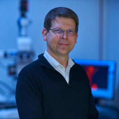
Kirk Czymmek, PhD
Director, Advanced Bioimaging Laboratory
Keith Duncan, FRMS
Research Scientist, Director of X-ray Imaging
David Huss, BS
Senior Laboratory Technician
Anastasiya (Nastia) Klebanovych, PhD
ABL Manager, Advanced Light Microscopy & Super-Resolution Imaging Expert
Lolita Rotkina, PhD
Staff Scientist III, SEM Specialist
Elizabeth VanHoecke, MSc
Research Associate, TEM Imaging Specialist
Phone
314.587.1261
Donald Danforth Plant Science Center
Advanced Bioimaging Lab
975 North Warson Road
St. Louis, MO 63132
Data Science