Down on the Farm
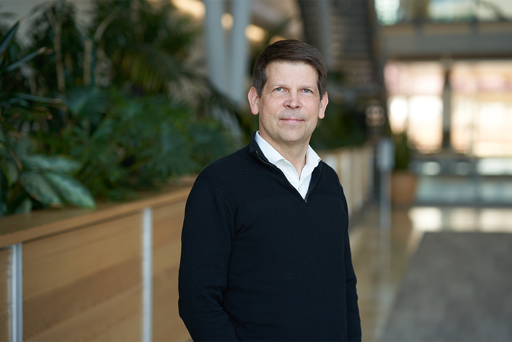
Growing up in the shadow of Finger Lakes Racetrack in upstate New York, Kirk Czymmek, PhD, learned a bit about horses, mostly what they eat.
The family grew cash crops, including alfalfa, timothy, and high-quality hay for the race horses. “As the middle child of seven on the farm, I was a laborer.” It was hot, hard work, but the free-range childhood inspired the young boy’s interest in science. Today, he has a PhD in plant pathology and is the director of the Danforth Center Advanced Bioimaging Laboratory.
Knowing the Unknown, Seeing the Invisible
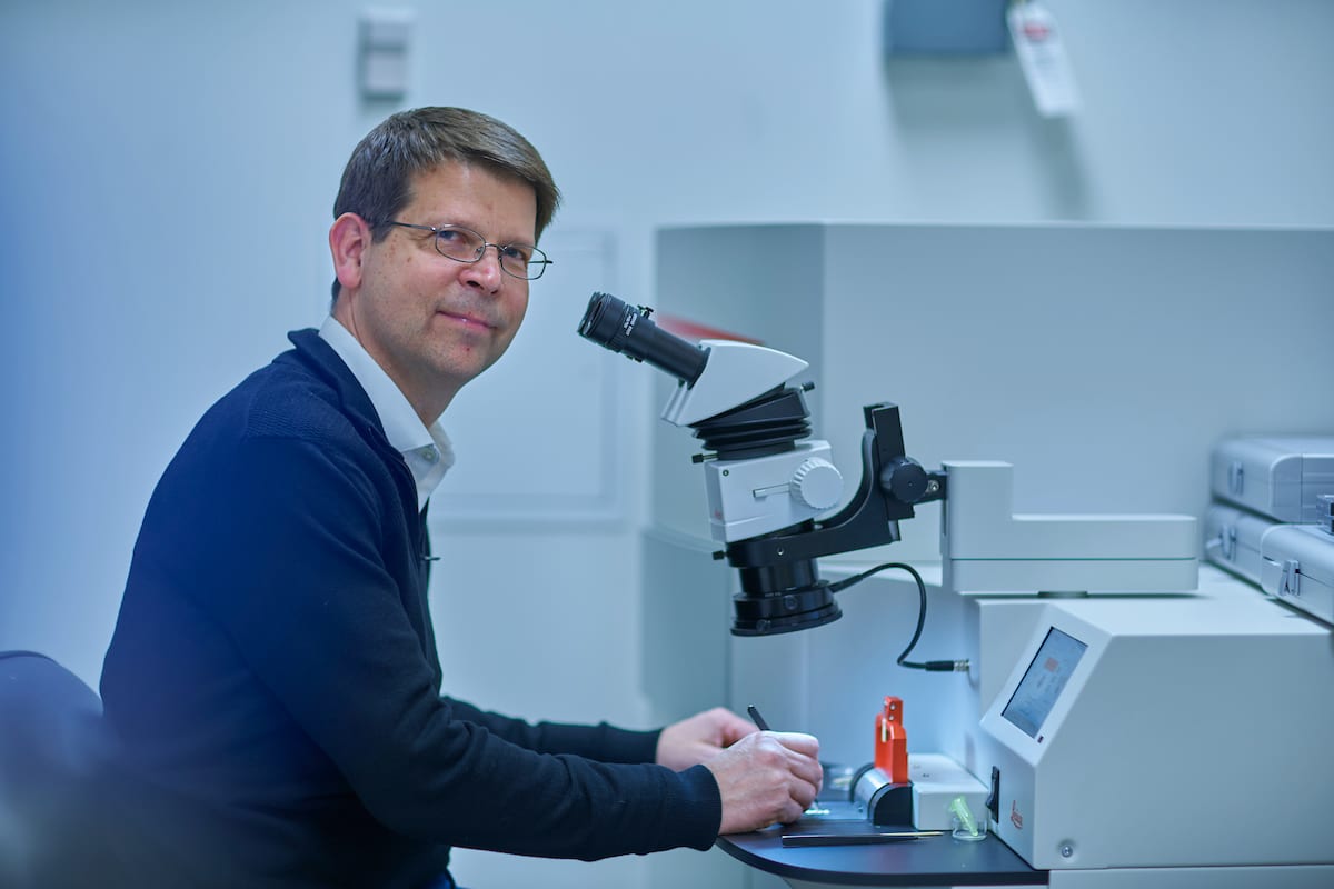
He remembers the exact moment he became interested in microscopes. “As an undergrad, I had an enthusiastic mentor who introduced me to microscopy. I was looking at these little cells growing at a very fast rate across the field-of-view through the microscope. They were filamentous fungi. You can actually see them growing, dividing and sporulating. The excitement of seeing the invisible struck me, and I knew this work could have significant impact on health and nutrition.”
Today, Kirk is an internationally renowned expert in bioimaging with 30 years’ experience and over 100 publications. He is proud of his role in discovering a new imaging approach to follow subcellular calcium signaling in filamentous fungi—a world first. His research today focuses on small microbes that cause disease in both humans and plants. And he is dedicated to his role at the Danforth Center, partnering with numerous colleagues to help advance their research as well.
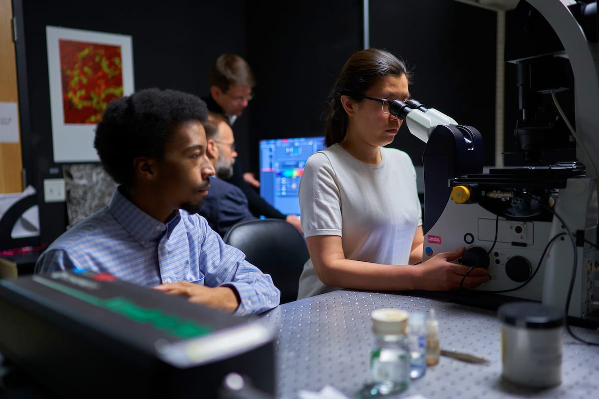
Best-in-Class Facilities
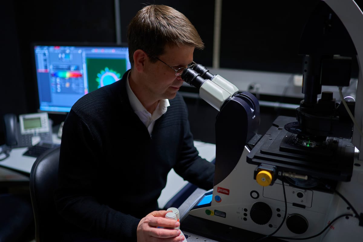
Today’s Advanced Bioimaging Lab is a far cry from the microscopy facilities of yore. Confocal microscopes are now generation 9. They are compact, but powerful, and allow researchers to look inside thick tissues and make revealing 3D perspectives. It’s kind of like an MRI scan, but at the cell or tissue level. The facility is adding new tech all the time, such as our recent addition of a highly automated 3D macro microsope that will really allow us to perform high-throughput live plant microscopy studies. Kirk adds: “Ultimately, our goal is to have the world’s best imaging capacity for plant research and become a center of collaboration far beyond the Danforth Center.”
Why He Gets Up in the Morning
“As the world’s warming continues, plants will likely be more stressed and more vulnerable. Under higher humidity and warmer environmental conditions, fungi and other pathogens will thrive and diseases once limited to the tropics will continue to spread. Danforth Center PIs are working to develop disease resistance in important plants - figuring out how to grow more food in challenging conditions. We need more productivity to feed the world. Advanced bioimaging will help.”
He adds: “The Danforth Center is a special place with a special mission. I get up every morning excited to do work that will benefit humanity.”
The Power of Peptides
In 2022, Kirk and his colleague Dilip Shah, PhD, developed a technology platform and co-founded Peptyde Bio. The startup was launched with the help of the Danforth Technology Company. Peptyde Bio discovers, designs, and characterizes novel anti-microbial peptides. These peptides can complement or replace chemical fungicides because they are effective, environmentally friendly, and have lower cost of development compared to traditional fungicides. With a strong pipeline developed through robust design capabilities, Peptyde Bio enables its partners to more quickly and cost effectively commercialize peptide-based biofungicides. Peptyde Bio was acquired by Invaio Sciences in December 2023.