Looking into the Future of St. Louis: Advanced Bioimaging
Would you like to see something unique?
At the Danforth Center, scientists utilize advanced technology to find solutions to some of the most critical problems facing our world.
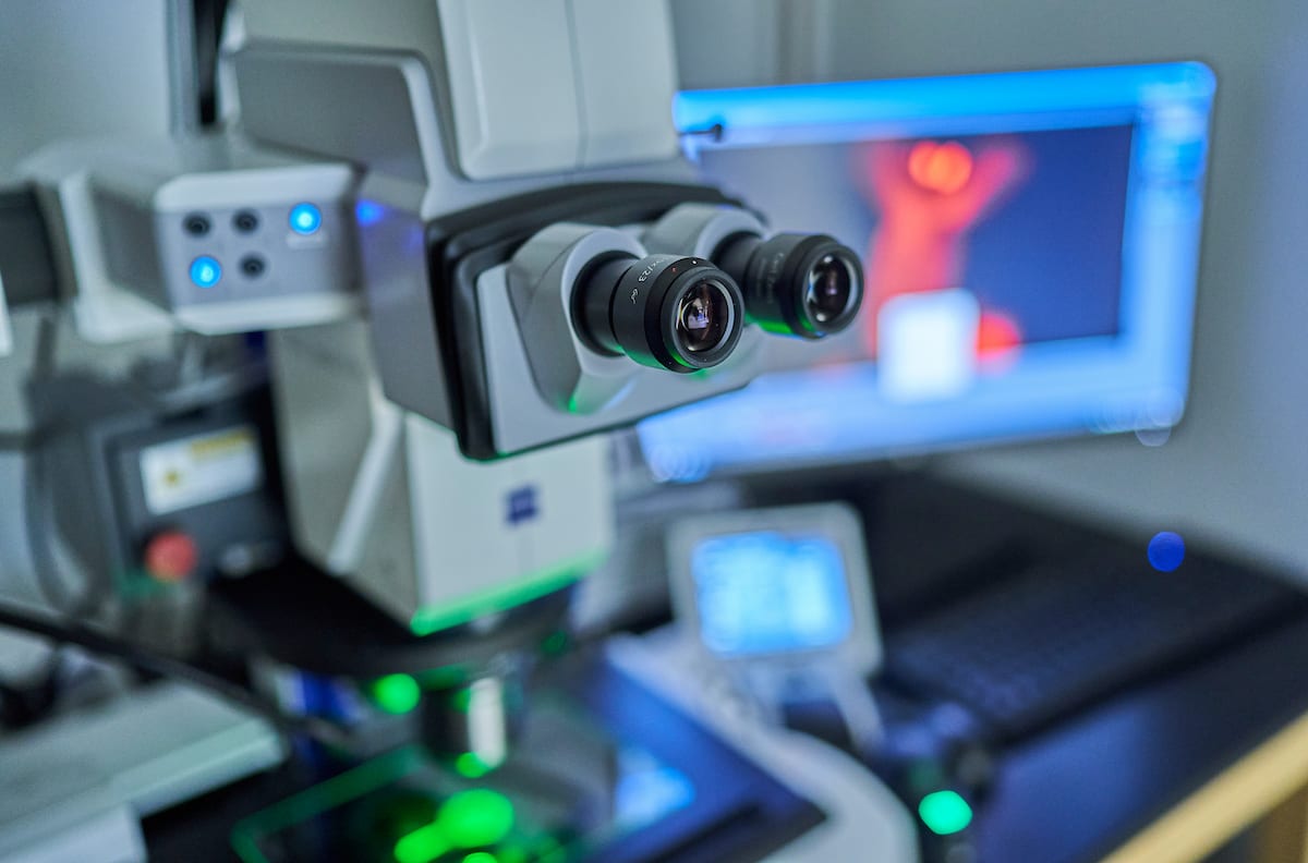
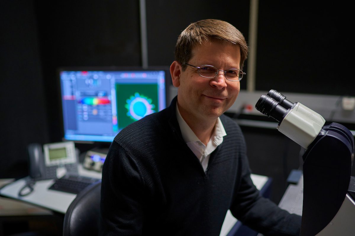
Much of this technology can be found in our core facilities, such as the Advanced Bioimaging Lab (ABL). Led by Director and Principal Investigator Kirk Czymmek, PhD, the lab uses state-of the art imaging technology to document plants, microbes, and their interactions from whole plants down to the cellular level. Recently, Dr. Czymmek showed us around the lab and gave us a brief overview of the work he and many other talented Center scientists do there. Watch the video below to see for yourself:
Seeing Potential
Because technology in optics and microscopy is constantly evolving, the ABL must constantly evolve as well. This means investing in the newest cutting-edge equipment. Just this year, the ABL added two new cutting-edge pieces, at a combined price tag of two million dollars. The purchase was made possible thanks to a Major Research Instrumentation grant from the National Science Foundation and to the generosity of individual donors to the Impact Fund.
The new ZEISS Elyra 7 super-resolution microscope, installed in January, combines laser light and a research microscope to create ultra-high-resolution and high-speed 3D imaging. This new scope can capture not only extremely tiny features, but also dynamic events in living and fixed cells. With super-resolution microscopy, scientists can see small cell molecules, structures, and organelles interact with each other and watch as a pathogen (virus, bacterium, fungus) invades and the plant defends itself.
The new ThermoFisher Scientific Talos L120C Transmission Electron Microscope (cryo-TEM) was installed this summer. Transmission electron microscopy (TEM), where a beam of electrons is transmitted through a specimen to form an image, is already cutting-edge technology. Cryo-TEM advances this approach one step further. It is a type of TEM where the sample is studied at room temperature and at cryogenic temperatures, approaching liquid-nitrogen-cooled temperatures of -145°C (-229°F) and below. This advanced platform is highly automated and has improved cryo capabilities (to image frozen samples) enabling us to visualize tiny plant structures, organelles and macromolecules in three dimensions at sub-nanometer resolutions in a more life-like state. It was purchased with the support of a generous donor.
These technologies and others in the ABL, along with the expertise of the ABL team, will allow Danforth Center researchers, as well as with partner companies and organizations, to discover more about the inner workings of plants than ever before.
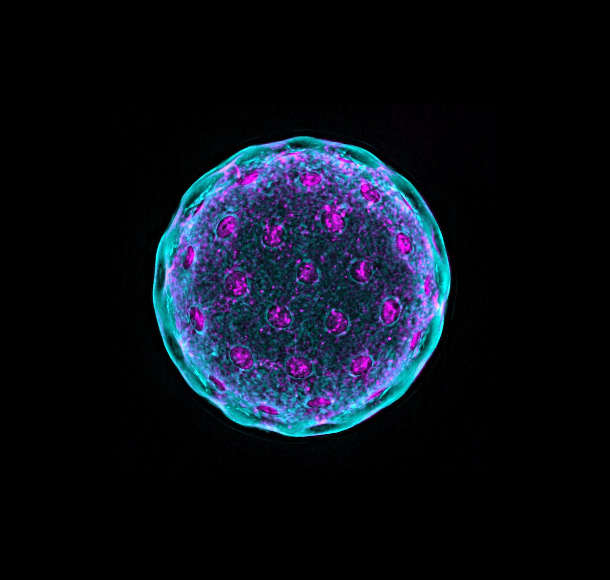
3D image of a quinoa pollen grain taken with the Danforth Center’s new super-resolution microscope, part of a study of heat stress by the Gehan lab. New technology like this is helping attract new business and innovation to St. Louis, while advancing the Danforth Center mission. Join us at danforthcenter.org/grow.
Already Advancing the Science
Since its arrival in January this year, the super-resolution microscope has been in high demand. This system has been used to study the microscopic effects of heat stress on quinoa pollen germination (Gehan lab, image above) and in model green algae Chlamydamonas (Zhang Lab), to witness the killing of fungal cells by small anti-microbial peptides derived from plants (Shah lab), to localize small RNAs in plant reproductive structures (Meyers lab), to understand the chemistry of the cell wall in the abscission zone of plants (Kellogg lab), and to multiplex label cell pathways in plant tissues (Czymmek lab).
The lab of Danforth Center Principal Investigator James Umen, PhD, studies fundamental biological questions such as cell size control and sex determination using green algae, including the protagonist of the video below: Chlamydomonas reinhardtii. Unlike in yeast and bacteria, where a mother cell divides once to generate two daughter cells, a C. reinhardtii cell has the potential to divide more than once to generate multiple daughters. See the lab’s award-nominated clip of this process below:
Image: “Hatchlings” This gif shows the hatching process of a green algal cell after three rounds of successive divisions, releasing eight daughter cells from the mother cell wall. The newborn daughters are leaving their siblings and starting to live their own lives. (Chloroplasts/chlorophyll are red. Nuclei are green. Cell walls are blue.) The video was taken by the ABL’s new super-resolution microscope with the help of Dr. Kirk Czymmek.
Having joined the Danforth Center August 1, Principal Investigator Tessa Burch-Smith, PhD, has already embraced the full suite of ABL equipment. Using the new cryo-TEM, Dr. Burch-Smith and her lab are able to create 3D reconstructions of intercellular channels at nanometer scale. This microscopy will help them answer key questions in plant cell biology. "This new microscope, along with some novel advanced sample preparation techniques being pioneered by Dr. Czymmek, should allow my lab to attain our goals. I’m very excited about joining the Danforth Center and having access to this cutting-edge technology and expertise."
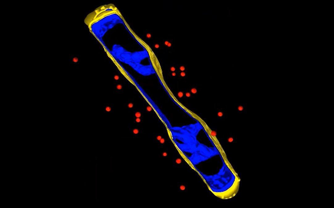
Video: A tomogram of two plasmodesmata (intercellular channels in plant cells). Traditionally, these 3D structures can only be viewed in two dimensions. But by using the Danforth Center’s new transmission electron microscope, Dr. Burch-Smith is able to view the structure as it truly is and gather more information than ever before. This in turn speeds her research that will help produce healthier and more nutritious crops. (Copyright American Society of Plant Biologists. www.plantcell.org)
Tools of the Trade
The ABL and the Danforth Center’s other core facilities do much more than groundbreaking scientific research. They are also bringing new investors, jobs, and opportunities to St. Louis. Through our state-of-the-art infrastructure, the Danforth Center campus attracts businesses from around the world to our region—and provides local startups access to the latest technology.
One company that is utilizing the unique resources of the Danforth Center ABL is Plastomics. Plastomics is developing a system that can introduce new traits into a plant via the chloroplast of cell, where photosynthesis occurs. Because this technology relies on cell parts, being able to look inside a plant cell and see the process in live time is crucial. Thanks to the Danforth Center and the new super-resolution microscope, scientists at Plastomics are able to do just that.
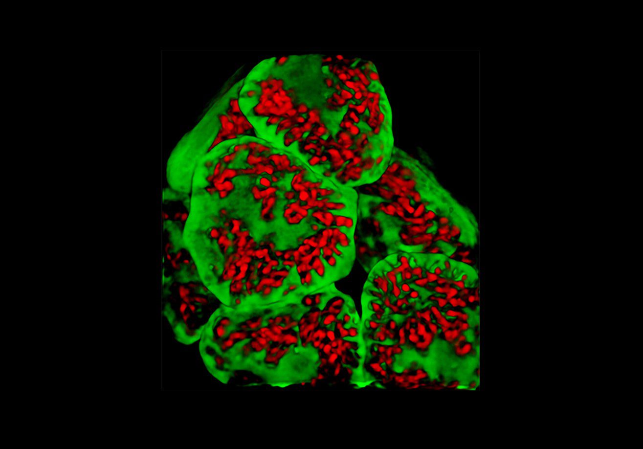
An image of soybean leaves taken with the Danforth Center’s new super-resolution microscope. The chloroplasts of these leaves are carrying a protein gene (green) that scientists at Plastomics had introduced into it. Being able to study this process using the Danforth Center’s unique facilities is crucial to the start-up company’s success.
“As a start-up company with limited resources, Plastomics has taken advantage of the cutting-edge technologies and facilities at the Danforth Center,” said Jeff Staub, the Founder CEO of Plastomics. “These facilities, which are unique in the region, provide resources and expert technical assistance that enables Plastomics to do research that would otherwise not be possible for us.”
Why It Matters
In recent years, a remarkable surge in new discoveries on the inner workings of cells have been forthcoming, benefiting from the rapid development of new genetic tools and cellular probes. At the same time, numerous and diverse novel imaging platforms have arisen, along with novel sample preparation methodologies leading a revolution in how a cells and tissues can be interrogated. This super-resolution microscope and cryo-TEM will advance existing Danforth Center imaging projects, catalyze new projects, and serve as a unique regional resource. Furthermore, this technology is advancing the ABL efforts to develop advanced imaging applications specifically targeted to plant science research.
The Danforth Center is helping to grow businesses in our region in many ways. Along with attracting outside companies with our state-of-the-art technology, the success of our work and our community continues to give rise to new, innovative spinout companies (like Benson Hill) dedicated to improving the world through the power of plant science. Generous donors to the Impact Fund help the Danforth Center acquire this leading-edge technology, and in turn, are helping transform the science… and our region.
Our goal is to have the world’s best imaging capacity for plant research and become a center of collaboration far beyond the Danforth Center.
-KIRK CZYMMEK, PhD, DIRECTOR, ADVANCED BIOIMAGING LABORATORY
Join Us in Building a Brighter Future
The Grow Challenge Week of Giving (Sept. 27 - Oct. 1) is an online peer-to-peer giving campaign, spearheaded by the Danforth Center Young Friends. Early donations are being accepted now. Join us as we build a brighter future. Click here to learn more or to donate.